Methods
Methods
X-Ray Reflectometry
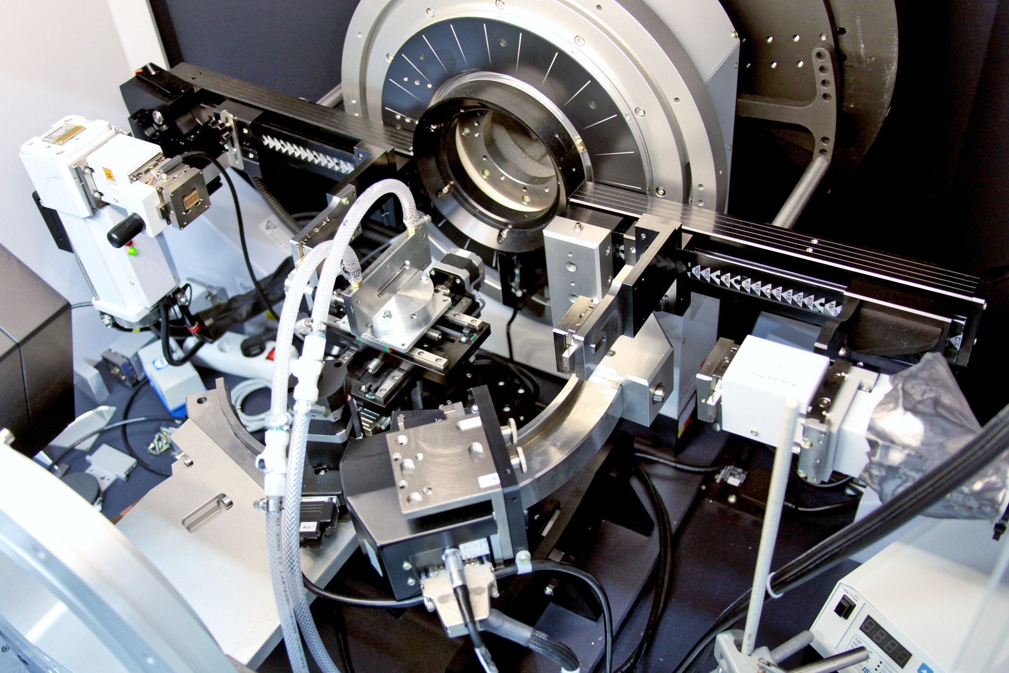
Rigaku Smartlab
Rigaku: Reflectometry, Diffraction and Reciprocal Space Mapping
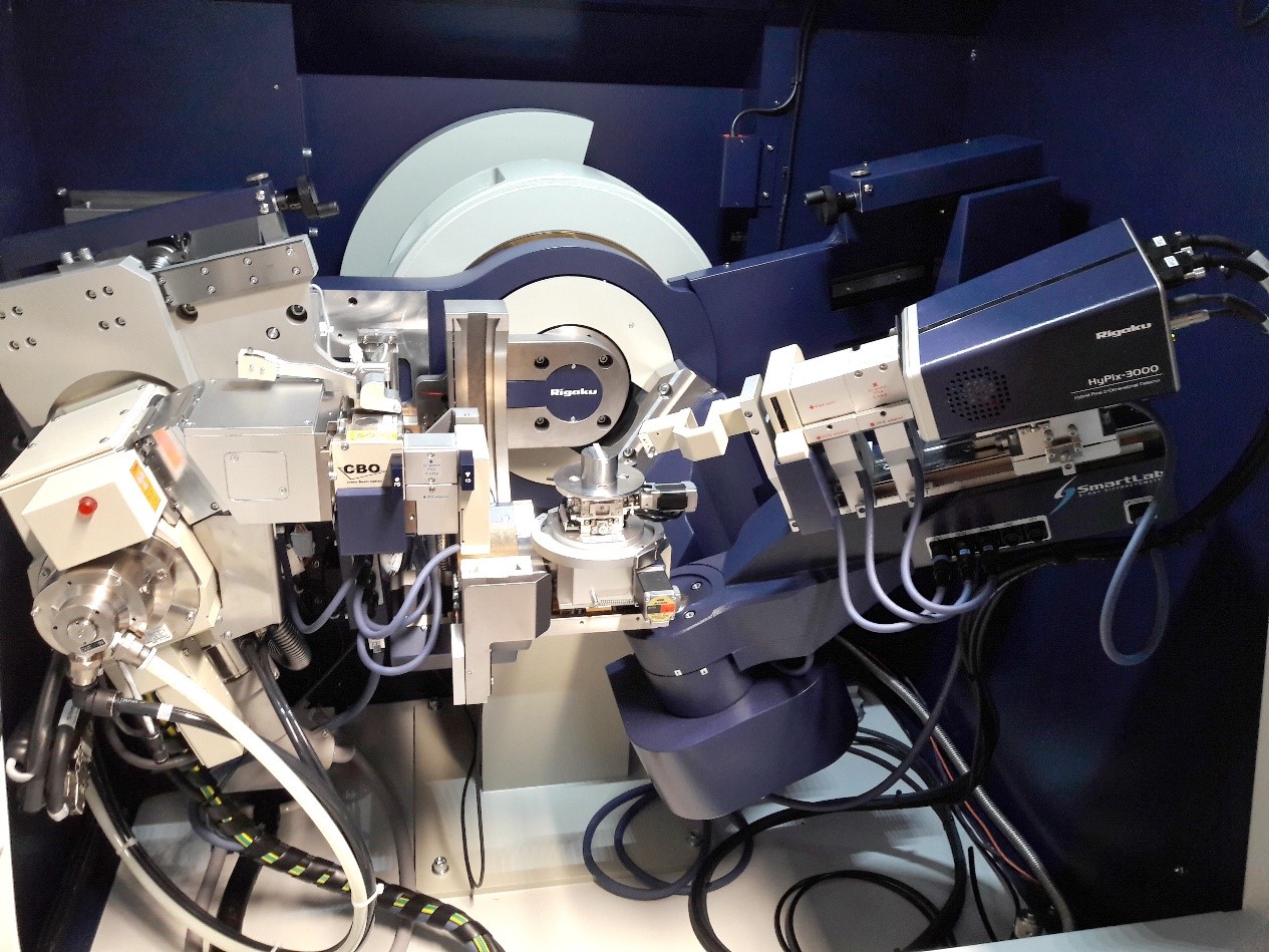
- Powder diffractometry with Q-Q-geometry (2Θ ca. -3° – 160°), gracing-incidence diffraction (GID), reflectometry
- X-ray source: rotating anode Cu (Kα), line or point focus system
- Running parameter 45kV, 160mA
- Filtering of Kβ, soller collimation, flexible absorber
- Primary optics: Goebel mirror, automated slit system, Johannsson monochromator and microfocus optics
- Sample environment: several different stages, Eulerian cradle
- Secondary optics: monochromator, automated slit system
- Detection system: Hypix 3000 solid state 2D-detector
- optional: capillary attachment, 0D- and 1D-detection system, heating equipment from Anton Paar
- Software: SmartLab Guidance, PDXL-Basic, PDXL-Qualitative, Global Fit XRR&RC
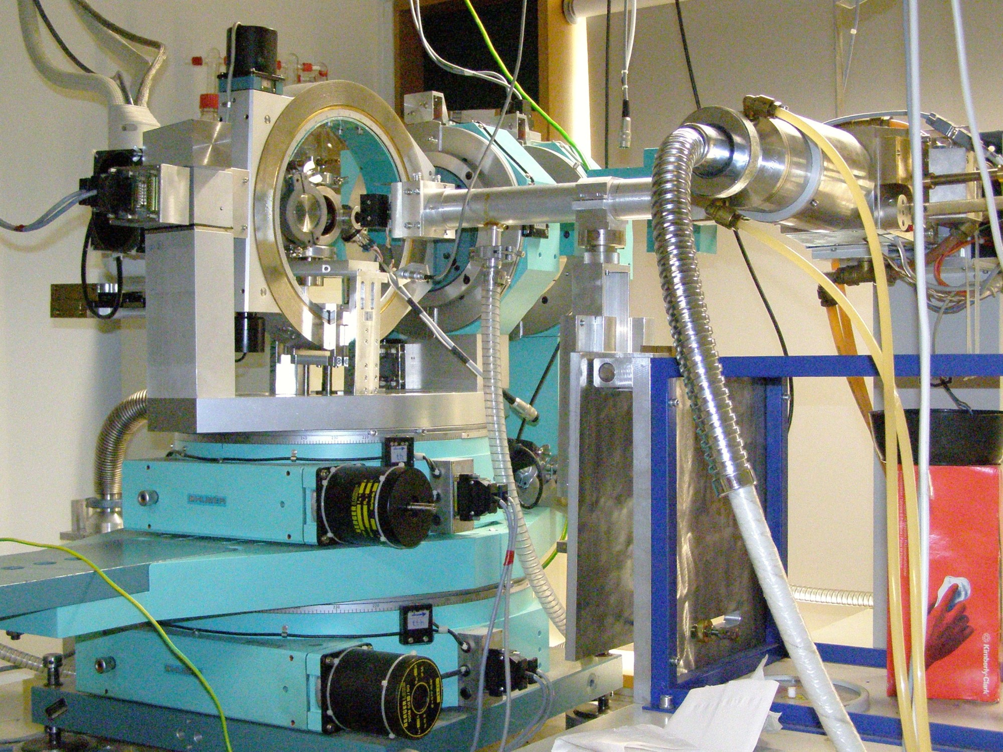
Bruker D8
Bruker D8 Discover for Reflectometry:
- Reflectometer with Q-Q-Geometry (2Θ ca. -10° – 140°) or Gracing-Incidence Diffraction (GID)
- X-ray source: Anode Cu (Kα) or Mo (Kα)
- Running Parameter 45kV, 45mA
- Filtering of Kβ, soller collimation, flexible absorber
- Primary optics: goebel mirror, slit system or asymetric channel cut monochromator (for both energies)
- Sample environment: several different stages, eulerian cradle
- Secondary optics: same as primary optics
- Detection system: 1-D semiconducting stripe detector (Lynxeye) or scintillation counter
- Optional: shear cell and heating equipment
Small Angle X-Ray Scattering
VAXSTER
VAXSTER
(Versatile Advanced X-ray Scattering Instrument Erlangen) for Small and Wide Angle Scattering
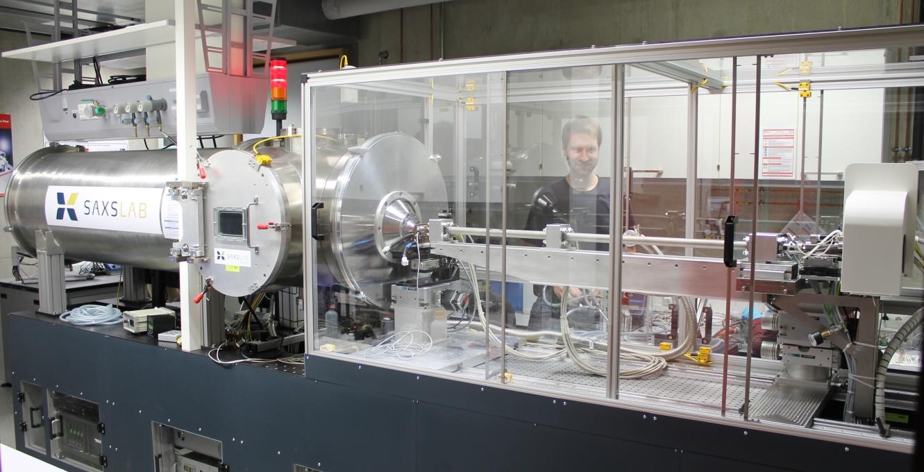
- Small- (SAX) and wide angle scattering (WAX), gracing incidence small angle scattering (GISAX)
- X-ray source: Excillum MetalJet, 10-70kV, focal spot size 5 – 20 mm^2@max. 250W, Ga (Kα, 1.34 A)
- Collimation: source multilayer optics, collimation through 3 sets of 4-bladed slits
- Sample stage: mounting of various large space sample stages inside detector tube possible (under vacuum ~10-5 bar), as well as externally (measurement in air/controlled vapor environment), yz-theta goniometer for transmission as well as grazing incidence applications
- Sample holders: Many different sample holders for mounting on yz-sample stage including: GISAXS sample holder, temperature-controlled Cu-holder with 5 sample-positions, Stopped-flow cell, capillary holder …
- Detection system: Pilatus 300K detector (500 Hz): three modules à 83.8×33.5 mm², 487×619 pixel à 172×172 µm², readout time of 3 ms, energy range of 4.5-36 keV at a resolution of 500 eV
- Q-range: Total Q-range: Q = 0.003-4Å-1
low count rates for small Q-limit, only achievable under ambient conditions;
high Q-limit only achievable in vacuum
individual configurations in between; e.g.: WAXS (0.05-2.5 Å-1), MAXS (0.012-0.67 Å-1), SAXS (0.006-0.26 Å-1), Extreme-SAXS (0.003-0.21 Å-1) - Software: SPEC
SAXSpace
SAXSpace (Anton Paar) for Small and Wide Angle Scattering
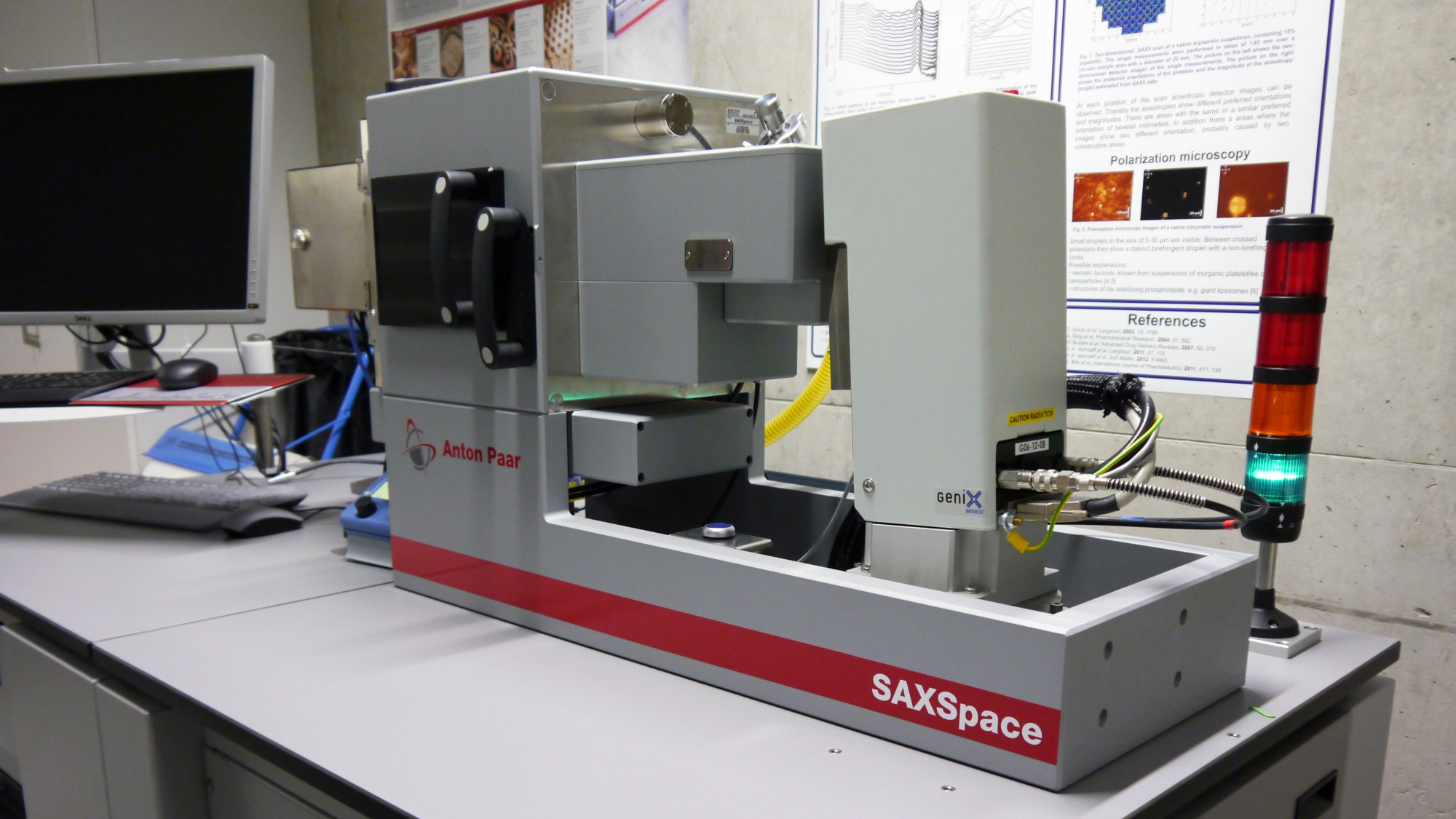
- Small angle (SAXS -> approx. 0.04° to 10°) and wide angle (WAXS, up to 74°) X-Ray scattering
- X-Ray source: 50 W Cu Kα microfocus tube
- Scatterless Kratky-base collimation system for line and point collimation
- Primary optics: focusing multilayer mirror to monochromatize and enhance X-Ray intensity
- Sample holders for liquids, pastes and solids
- Detection system: 2-D diode-array (CMOS) detector (Pilatus 100k)
- Temperature range of -30°C to +150°C (peltier heating/cooling)
- Software: SAXSdrive, SAXStreat and SAXSquant
High Energy X-Ray Diffraction
High Energy X-Ray Laboratory HEXBay

- Focussing Laue method (reflection topography, focussing distance up to 8m, resolution 0.005°), powder diffraction, real structure analysis
- Motorized collimation system with tungsten blades
- Sources: 2 tungsten tubes with
- up to 450 KV, 2.5 mm (0.9 kW) / 5.5 mm (4.5 kW) focal spot (EN 12543)
- up to 225 KV, 0.4 mm (0.8 kW) / 1 mm (1.8 kW) focal spot (EN 12543)
- Sample stage: heavy load sample stage (up to 150 kg), Eulerian cradle, omega stage with resolution of 0.2“
- Sample environment: several furnaces (T up to 1800°C, vacuum, oxygen or inert gas environment), pressure system
- Detection systems:
- Imageplate MAR 345, 345 cm diameter, max. resolution 100µm, readout time at max. resolution ~2 min
- 2 D scintillation system, active area 30 cm x 20 cm, 2 x 14 Bit peltier cooled CCD with 1280 x 1024 pixel, resolution ~ 150 µm, readout time 125 ms
- Software: SPEC
X-Ray Powder and Single Crystal Diffraction
Philips/PANalytical X´Pert Pro-MPD Powder Diffractometer
Philips/PANalytical X’Pert Pro-MPD Powder Diffractometer

- Powder diffraction with Q-Q-Bragg-Brentano geometry (2Θ ca. 5° – 100°), gracing-incidence powder diffraction (GID, incidence angle>≈0.2°), reflectometry
- X-ray source: Anode Cu (Kα)
- Running Parameter 40kV, 35mA
- Filtering of Kβ, soller collimation
- Primary optics: Goebel mirror, slit system
- Sample environment: various sample holder systems as reflectometry stage, Eulerian Cradle …
- Detection system: semiconducting stripe detector (X’Celerator) or Gas ionisation counter (Miniprop)
- Variable step size
- optional: capillary optics
Huber Guinier Diffractometer
Huber Guinier Diffractometer

- Powder diffraction in transmission-geometry (2Θ fixed 10° – 100°)
- X-ray source: Anode Cu (Kα)
- Running Parameter 40kV, 35mA Primary optics: monochromator: Kα1, high angular resolution
- Detection system: Image plate online read-out, illumination time: ca. 10min – several hours
- Software: Huber guinier software
Rotating Anode Setup for Powder Diffraction
Rotating Anode Setup for Powder Diffraction:
- Powder measurement in transmission Debye-Scherrer geometry, texture analysis
- X-ray source: rotating anode Cu (Kα)
- Voltage and current up to 44 kV and 75 mA, respectively
- Primary optics: crossed Goebel mirror setup
- Sample environment: 6 axis goniometer for adjustment of sample position, In-Situ chamber for variation of temperature ramps up to 550° C, possibility for different atmospheres (vaccum, inert gases)
- Detection system: 2D ccd detector allows measurement of full diffraction rings (2-Q range from 0 -50°)
- Software: SPEC
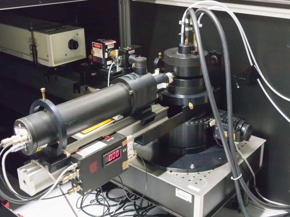
Huber Laue System
Huber Laue System

- Laue Camera for transmission- and backscattering diffraction
- Anode Molybdenum
- Image plate detection
- Primary optics: different collimation systems
Light Scattering and Spectroscopy
BI-200SM Goniometer (Brookhaven Instruments Corporation)
BI-200SM Goniometer (Brookhaven Instruments Corporation)
- motor driven angle selection (0.01° steps)
- narrow-band optical filters for laser wavelegths of 633, 514.5, 488 nm
- 100, 200, 400 μ pinholes for selecting coherence areas (QELS measurements)
- 1, 2, 3mm apertures for adjusting intensity (classical measurements)
- Selected photomultiplier tube (PMT) as Detector
- circulating index matching liquid with dust filter
- Temperature control approximately+5°C to 80°C
- needed sample Volume: 1.5mL (strongly diluted)
- Diode Laser 637 nm (BIC), 30mW
- Lexel95-2 (Lexel), Argon Laser, 476.5 nm – 514.5 nm, 4 W, 1.33 mm beam diameter, 0.6 mrad beam divergence
UV-Vis Spectrometer
UV-Vis Spectrometer TIDAS S 500K
(TIDAS S 500K UV/NIR 1910 DH, J&M Analytik AG)

- wavelength range 187.79 – 1016.53 nm
- spectral resolution < 2.5 nm
- wavelength accuracy < 1.0 nm
- wavelength reproducibility 0.1 nm
- baseline drift at 250 nm: 5 μAU/h
- signal-to-noise ratio < 20 μAU
- integration-time range 0.7 – 10 000.0 ms
- integrated D2- & halogen light source
- detector MCS – Aspen 780 kHz
- photodiode array: 1024 pixels
- Bio-Kine 32 V4.66 for data acquisition and analysis
Thermoanalysis
Micro DSC III
Micro DSC III Analyzer (Setaram)

- High sensitivity DSC and calorimetry
- Temperature range: -20 to 120 °C
- Scanning rate: 0.001 to 1.2 °C/min
- Vessel volume: 850 µl
- Resolution: 0.3 µW
- Cooling rate from 120°C to room
- temperature: 1.5 h
- Scanning or isothermal mode
- Thermal Analysis Software: Calisto
DSC 8500
Perkin Elmer DSC 8500 (Perkin Elmer)
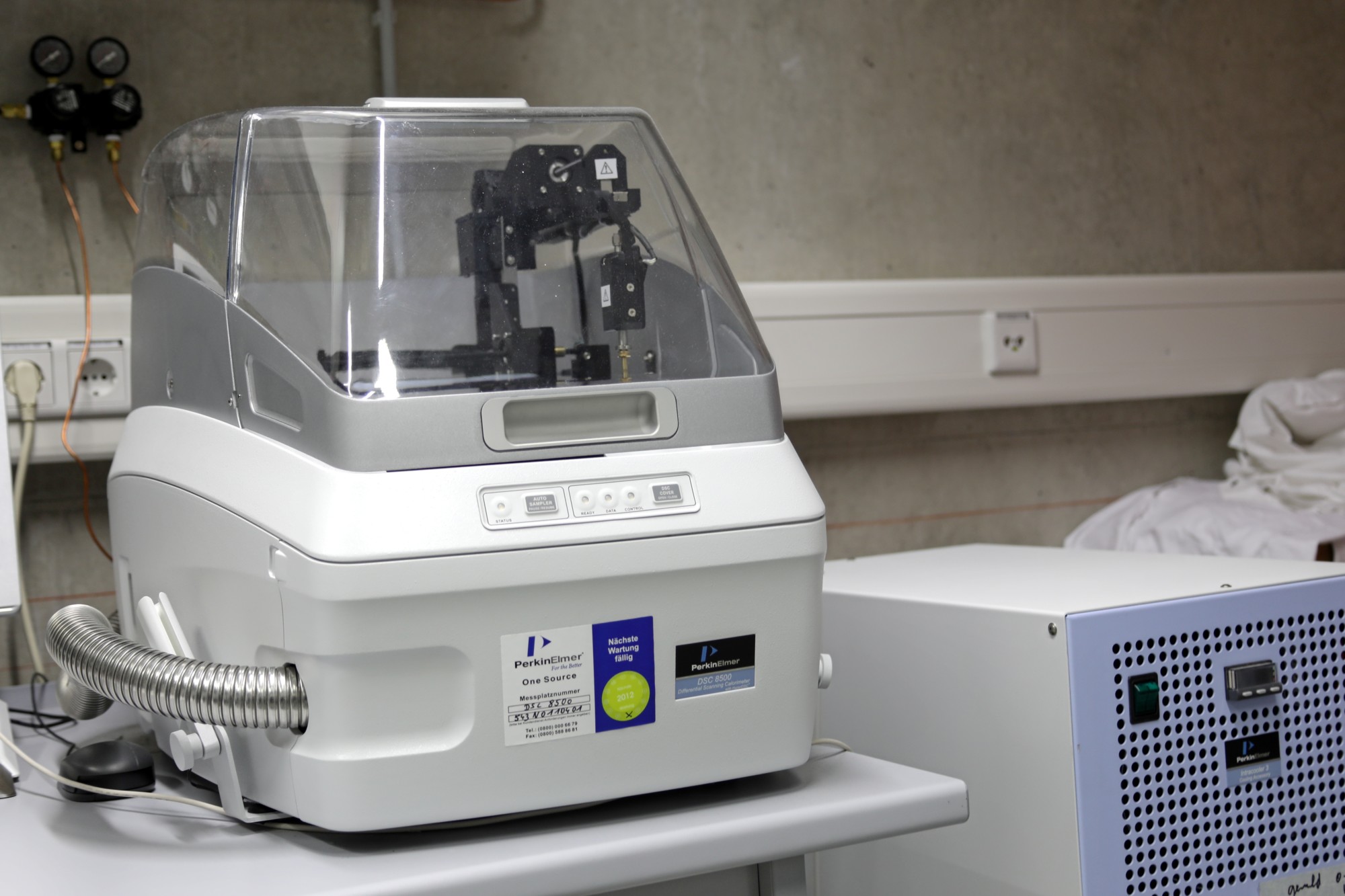
- Hyper-enabled Double-Furnace Differential Scanning Calorimeter
- Cooling system: Intracooler 3 (Perkin Elmer), three stage, closed-loop circulationg heat exchanger
- Temperature range: -100 to 750 °C (by nitrogen usage)
- In-situ ballistic cooling to 2100 °C/min
- Scanning rate: 0.1 to 750 °C/min
- Sample pans: aluminium (50 µl)
- Analysis Software: Pyris
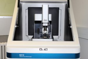
Atomic Force Microscope
Park Systems NX10 AFM
Z ScannerGuided high-force flexure scanner
|
XY ScannerSingle module flexure XY-scanner with closed-loop control |
StageZ stage range : 25 mm
|
Vision10x (0.21NA) ultra-long working distance lens (1µm resolution) |
SoftwareSmartScan™Dedicated system control and data acquisition software XEIAFM data analysis software
|
Standard ImagingTrue Non-Contact AFM Force MeasurementForce Distance (FD) Spectroscopy |
Special environment
Stopped-Flow SFM-2000
(SFM-2000, Bio-Logic)

- two 10 ml syringes
- minimal injection volume per syringe: 28 μl
- flow rate from 0.062 – 10 ml/s per syringe
- minimum total flow rate for efficient mixing: 1 ml/s
- ratio range from 1:1 to 1:40
- duration of flow: 1 ms to 60 000 ms per phase
- minimal dead time 0.7 ms
- Berger ball mixer
- Bio-Kine 32 V4.66 for syringe control